Home / Applications / Preclinical research

Measurement technology for preclinical research

Preclinical researchers often face data collection challenges, like accuracy, repeatability, the need to translate data to clinical applications, and measurement tools that inadvertently introduce artifacts or stress responses in the models. Overcoming these challenges requires precise, minimally invasive tools that can capture the subtle biomarkers of disease. PeriCam PSI is a noninvasive, non-contact, and non-destructive blood perfusion imaging system that delivers and visualizes robust macro- and microcirculation insights, providing the capabilities to record and observe the effect of treatments and provocations in real-time.

Why measure microcirculation?

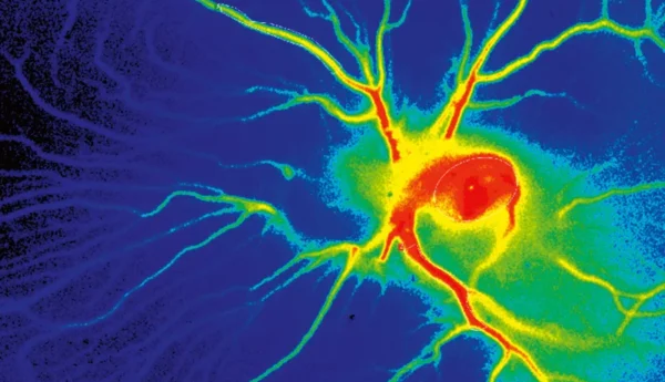
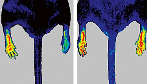
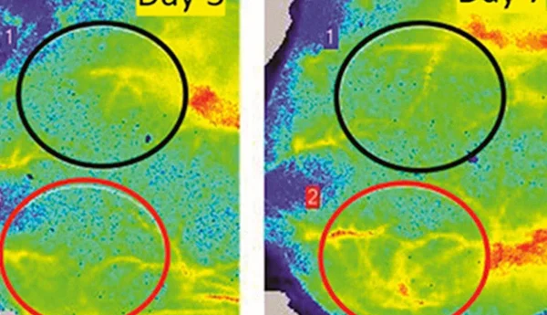
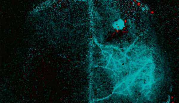
Measuring microcirculation in preclinical research is useful because this is where critical biological processes like nutrient delivery, waste removal, and immune response occur. Early signs of disease, such as impaired blood flow in diabetes or inflammation in cardiovascular conditions, often appear at the microvascular level. By closely monitoring microcirculation in preclinical models, researchers can detect these early changes, evaluate the effects of interventions, and study disease progression in a way that provides valuable insights for potential clinical applications.

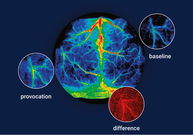
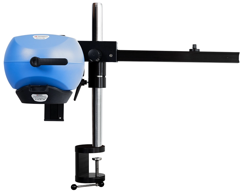
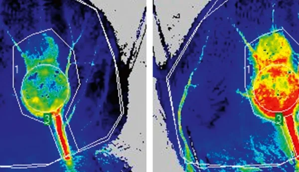
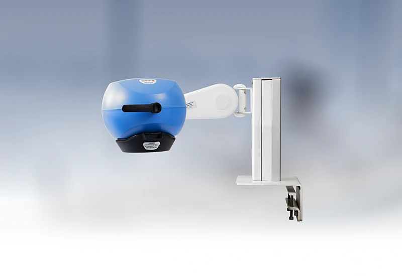
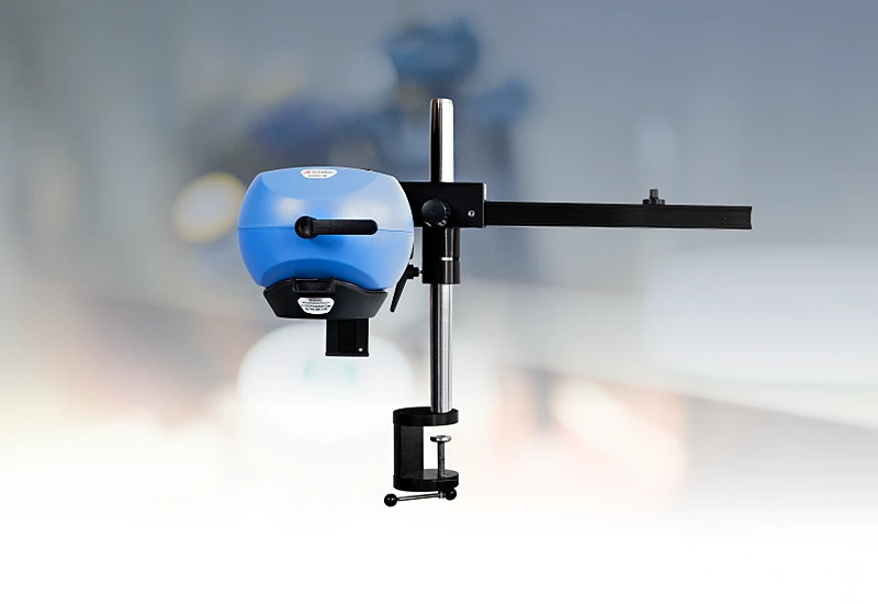
PeriCam PSI leverages laser speckle contrast imaging (LSCI) to assess blood perfusion in tissue at the microcirculation level with detail and precision. To meet the demands posed by highly diverse research — and clinical — settings, we’ve designed several PeriCam PSI models. PeriCam PSI HR (high resolution) works from a fixed distance and is specifically equipped to measure small animals such as rats and mice. For environments that demand versatility, PeriCam PSI NR supports a range of working distances. Both models can be upgraded with zoom functionality, combining high resolution and flexibility in a single instrument.

Features

Background image
  • Rapid data analysis, delivering perfusion graphs, images, and video recordings in real-time.
  • Regions of interest (ROIs) and time periods of interest (TOIs) that can be defined and updated before, during, and after recording.
  • Frequency of up to ~100 images per second.
  • Automated background compensation for robust measurements under varying lighting conditions.
  • Suitable for longitudinal studies.
  • High-quality arm with good maneuverability to ease positioning, and stability to prevent movement artifacts. 
  • Wide-angled color camera for documentation of the measurement area and its surroundings, easing post-measurement analysis.
  • Non-contact, non-destructive, and noninvasive

References

  1. Creager, M. A., Matsushita, K., Arya, S., Beckman, J. A., Duval, S., Goodney, P. P., Gutierrez, J. A. T., Kaufman, J. A., Joynt Maddox, K. E., Pollak, A. W., Pradhan, A. D., & Whitsel, L. P. (2021). Reducing Nontraumatic Lower-Extremity Amputations by 20% by 2030: Time to Get to Our Feet: A Policy Statement From the American Heart Association. Circulation, 143(17), e875–e891. https://doi.org/10.1161/CIR.0000000000000967
  2. Zhang, P., Lu, J., Jing, Y., Tang, S., Zhu, D., & Bi, Y. (2017). Global epidemiology of diabetic foot ulceration: a systematic review and meta-analysis Annals of medicine49(2), 106–116.
  3. Settembre, N., Kagayama, T., Kauhanen, P., Vikatmaa, P., Inoue, Y., & Venermo, M. (2018). The Influence of Heating on Toe pressure in Patients with Peripheral Arterial Disease. Scandinavian journal of surgery : SJS : official organ for the Finnish Surgical Society and the Scandinavian Surgical Society107(1), 62–67. https://doi.org/10.1177/1457496917705994