Cutaneous wound healing
To support the metabolic needs of tissue repair during wound healing, the formation of angiogenic capillaries and their organization into microvascular networks is essential. Diseases like diabetes disrupt this process, leading to wound-healing impairment. Owing to the global prevalence of this issue, the development of materials and therapeutics to promote wound healing is a major area of research.

Data collection challenges
Researchers often use the skin as a model for testing new treatments, such as topical drugs, dressings, biomaterials, and tissue-engineering techniques. Cutaneous wound healing can be used to evaluate how interventions influence the healing process and their potential to enhance wound closure or reduce scar formation.
Following wound creation, subjects are measured at various intervals during the two-week healing period. Typically, researchers look for reductions in wound size and mature vasculature formation using CD31 and α-SMA immunohistochemistry. However, this approach involves euthanizing the animals at each measurement.
Overcoming challenges with PeriCam PSI
PeriCam PSI can measure blood perfusion in wounds over the entire two-week healing period on the same animal, reducing the number of subjects needed and supporting robust findings. The data can be used to evaluate pro-angiogenic effects of therapeutic agents or natural recovery in healthy compared with diseased (diabetic) and wild-type contra knockout animals [1] [2] [3] [4].
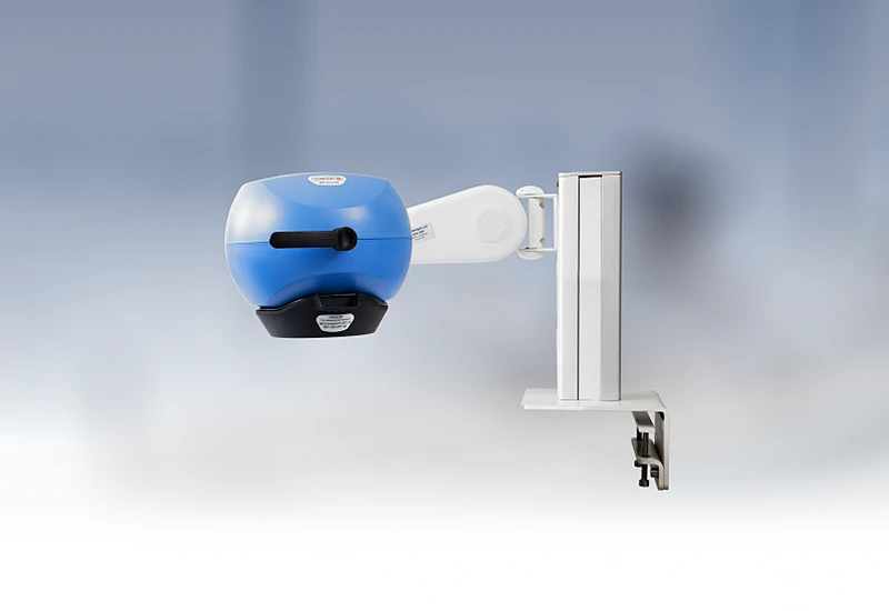
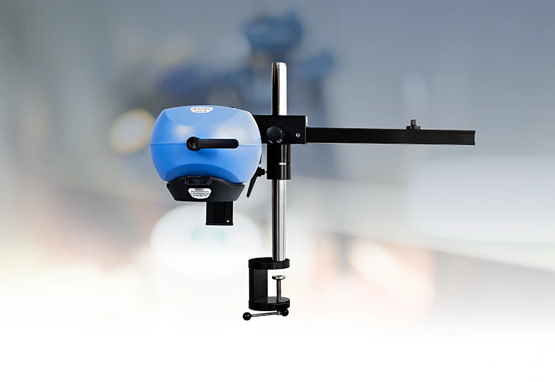
PeriCam PSI HR

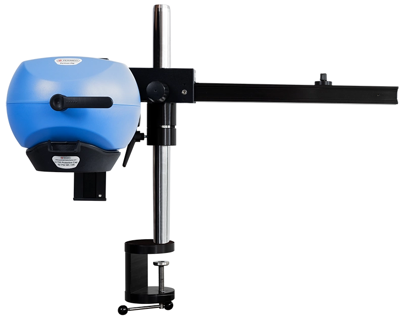
PeriCam PSI leverages laser speckle contrast imaging (LSCI) to assess blood perfusion in tissue at the microcirculation level with detail and precision. PeriCam PSI HR (high resolution) works from a fixed distance and is specifically equipped to measure small animals such as rats and mice.
- Rapid data analysis, delivering perfusion graphs, images, and video recordings in real-time.
- Regions of interest (ROIs) and time periods of interest (TOIs) that can be defined and updated before, during, and after recording.
- Frequency of up to ~100 images per second.
- Automated background compensation for robust measurements under varying lighting conditions.
- Suitable for longitudinal studies.
- High-quality arm with good maneuverability to ease positioning, and stability to prevent movement artifacts.
- Wide-angled color camera for documentation of the measurement area and its surroundings, easing post-measurement analysis.
- Non-contact, non-destructive, and noninvasive.
Related products
Contact us
Get in touch
For more information about PeriCam PSI and how it can help you with CBF preclinical studies, fill out the form and we will be in touch shortly.
References
- Cutaneous Epithelial to Mesenchymal Transition Activator ZEB1 Regulates Wound Angiogenesis and Closure in a Glycemic Status–Dependent Manner. Singh, Kanhaiya and Sinha, Mithun and Pal, Durba and Tabasum, Saba and Gnyawali, Surya C. and Khona, Dolly and Sarkar, Subendu and Mohanty, Sujit K. and Soto-Gonzalez, Fidel and Khanna, Savita and Roy, Sashwati and Sen, Chandan K. 11, 2019, Diabetes, Vol. 68, pp. 2175-2190.
- Correction of MFG-E8 Resolves Inflammation and Promotes Cutaneous Wound Healing in Diabetes. A. Das, S. Ghatak, M. Sinha, S. Chaffee, Noha S. Ahmed, N. L. Parinandi, E. S. Wohleb, J. F. Sheridan, C. K. Sen, and S. Roy. 12, 2016, The Journal of Immunology, Vol. 196, pp. 5089-5100.
- Circulating Exosomal miR-20b-5p Inhibition Restores Wnt9b Signaling and Reverses Diabetes-Associated Impaired Wound Healing. Yuan Xiong, Lang Chen, Chenchen Yan, Wu Zhou, Yori Endo, Jing Liu, Liangcong Hu,Yiqiang Hu, Bobin Mi and Guohui Liu. 3, 2020, Small, Vol. 16, p. 1904044.
- Saliva Exosomes-Derived UBE2O Promotes Angiogenesis in Cutaneous Wounds by Targeting SMAD6. Bobin Mi, Lang Chen, Yuan Xiong, Chenchen Yan, Hang Xue, Adriana C. Panay, Jing Liu, Liangcong Hu, Yiqiang Hu, Yun Sun, Faqi Cao,
LASCA and LSCI
Laser speckle contrast imaging (LSCI) is the innovative technology behind PeriCam PSI, offering non-contact, high-resolution perfusion imaging. The technique was first described by J.D. Briers and S. Webster in 1996 and referred to as laser speckle contrast analysis (LASCA). In later years, the term LSCI gained in popularity and is nowadays the more common term. However, LSCI and LASCA both refer to the same technique, providing precise and reliable perfusion data without the need for contrast agents.