Subcutaneous tumor model
The subcutaneous tumor model is a popular assessment system for in vivo evaluation of cancer medicine candidates. This model often uses immunodeficient animal strains, where cultured cancer cells are implanted subcutaneously, taking about two weeks to produce a solid tumor.
The growth and metastasis of cancer tumors rely on the development of blood vessels and an effective microcirculation. Without angiogenesis, a small focus of tumor cells cannot grow at a secondary site. As such, angiogenesis plays a central role in tumor growth, progression, invasion, and metastasis. Inhibiting angiogenesis provides a potential strategy for some treatments.
Because it can measure and visualize blood perfusion in and around subcutaneous tumors over time, PeriCam PSI can help researchers monitor tumor progression and validate the efficacy of anti-angiogenic therapies for cancer treatment.
Overcoming data collection challenges
Tumor tissue is dynamic, undergoing rapid changes in blood flow, hypoxia, and metabolic activity. Capturing these changes requires real-time, high-resolution data acquisition. PeriCam PSI addresses this challenge by supporting continuous, noninvasive monitoring.
PeriCam PSI measures blood perfusion in a noninvasive, non-contact, and non-destructive way. This design approach supports longitudinal monitoring of angiogenesis without disruption to the tissue.
While measurement tools cannot eliminate data variability caused by biological differences in test subjects and experimental conditions, they can help ensure that additional variability is not introduced into the results. PeriCam PSI is designed to deliver robust measurements — objective, repeatable, precise, and accurate — helping to minimize additional variability and enable cross-study comparisons.
PeriCam PSI in practice
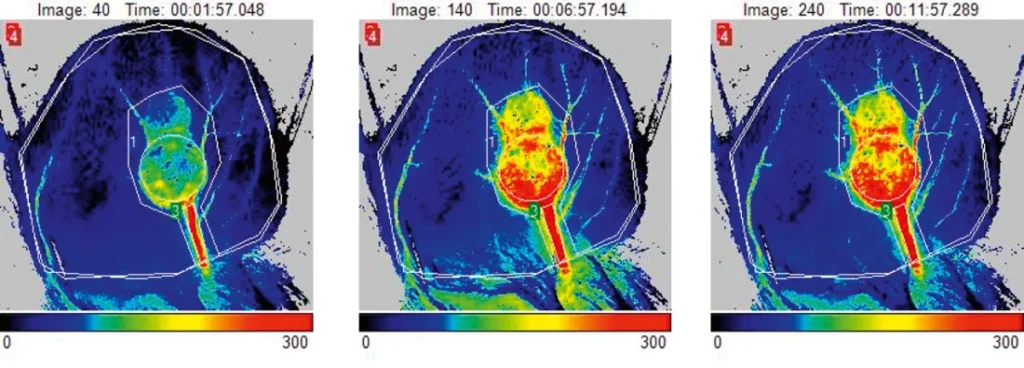
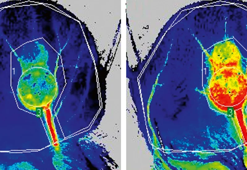
These perfusion images generated by PeriCam PSI show a mouse ear with a subcutaneous tumor on day-14. The images show how blood perfusion in the tumor increases in response to a heat challenge.
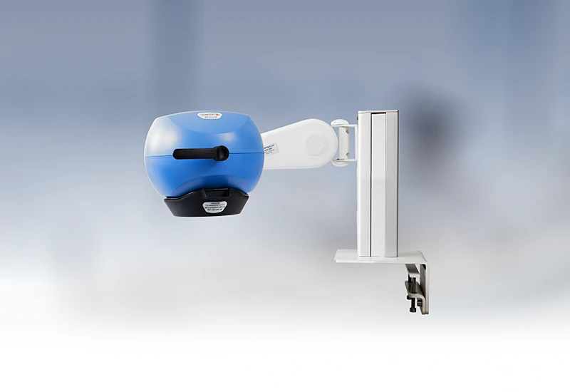
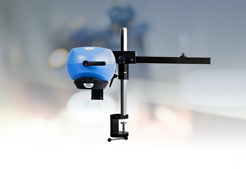
PeriCam PSI HR

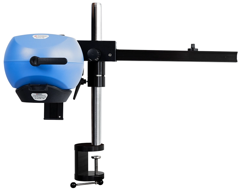
PeriCam PSI leverages laser speckle contrast imaging (LSCI) to assess blood perfusion in tissue at the microcirculation level with detail and precision. PeriCam PSI HR (high resolution) works from a fixed distance and is specifically equipped to measure small animals such as rats and mice.
- Rapid data analysis, delivering perfusion graphs, images, and video recordings in real-time.
- Regions of interest (ROIs) and time periods of interest (TOIs) that can be defined and updated before, during, and after recording.
- Frequency of up to ~100 images per second.
- Automated background compensation for robust measurements under varying lighting conditions.
- Suitable for longitudinal studies.
- High-quality arm with good maneuverability to ease positioning, and stability to prevent movement artifacts.
- Wide-angled color camera for documentation of the measurement area and its surroundings, easing post-measurement analysis.
- Non-contact, non-destructive, and noninvasive.
Related products
Contact us
Get in touch
For more information about PeriCam PSI and how you can use it to overcome the data collection challenges associated with preclinical research, fill out the form and we will be in touch with you shortly.
LASCA and LSCI
Laser speckle contrast imaging (LSCI) is the innovative technology behind PeriCam PSI, offering non-contact, high-resolution perfusion imaging. The technique was first described by J.D. Briers and S. Webster in 1996 and referred to as laser speckle contrast analysis (LASCA). In later years, the term LSCI gained in popularity and is nowadays the more common term. However, LSCI and LASCA both refer to the same technique, providing precise and reliable perfusion data without the need for contrast agents.