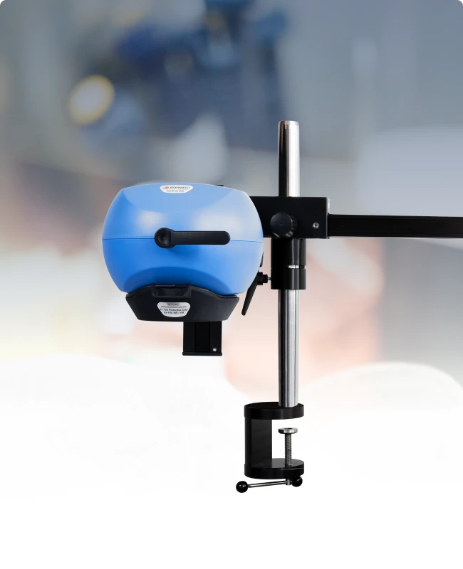
PeriCam PSI HR
Real-time perfusion imaging for research
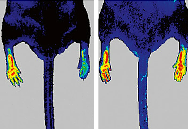
Primarily designed for preclinical research, PeriCam PSI HR leverages laser speckle contrast imaging (LSCI) to measure blood perfusion in tissue at the microcirculation level with precision in real-time. The HR model is specifically equipped to measure small animals, such as rats and mice, in a non-destructive way — without contact, invasive probes, or tracer agents.

Data and images
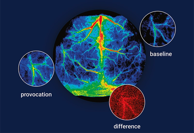
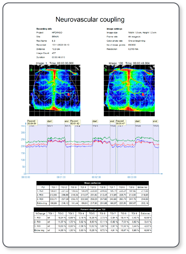
PeriCam PSI HR converts blood perfusion in tissue into a continuous, real-time data stream of perfusion units. To simplify interpretation, the application software (PIMSoft) translates these measurements into clear, color-coded images and graphs.
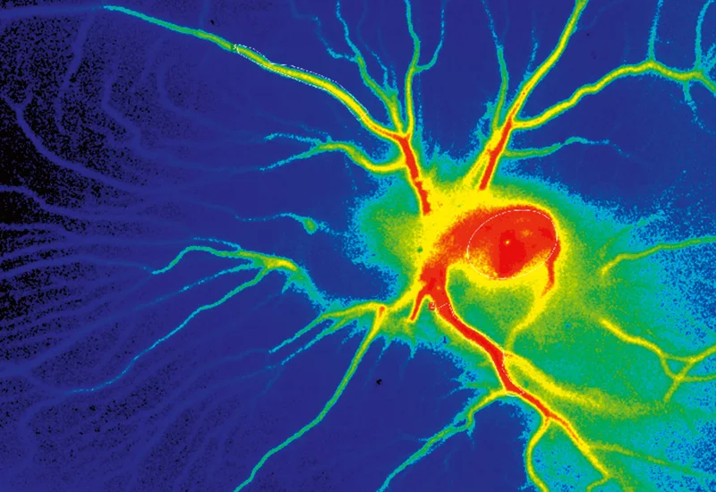
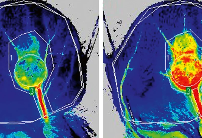
The color coding in these mouse brain perfusion images, for example, is set to the default, where blue and cyan indicate the lowest levels of perfusion, orange and red the highest, and green and yellow in between.
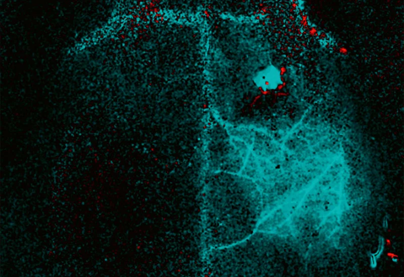
In the experiment, researchers stimulate the whiskers on the right side of the mouse’s head. The baseline and provocation images show the effect on blood perfusion on the left side of the brain. For easy comparison, PeriCam PSI HR provides difference images visualizing the change in perfusion between the baseline and provocation. Images are color-coded on a red-to-black-to-cyan scale: shades of red indicate increased perfusion from baseline to provocation, black represents no change, and shades of blue indicate decreased perfusion.
High resolution
Adapted for preclinical research where high resolution is desired to reveal details, PeriCam PSI HR works at a fixed distance of 13 cm and offers magnification optics of 10 µm/pixel for up to 2.1 x 2.5 cm. With zoom functionality, measurement areas can be up to 20 x 24 cm.
Zoom functionality unlocks the camera optics to expand the measurement flexibility of your instrument and broadens its field of application.

Features
Regions of interest (ROIs)

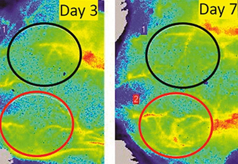
To support targeted analysis, PeriCam PSI HR allows you to define regions of interest (ROIs). This feature facilitates observation of localized changes in perfusion in response to stimuli or interventions in specific areas, such as a part of the brain or tissue. ROIs can be customized in shape and size within PIMSoft, enabling precise and repeatable measurements for consistent results across experiments.
Time periods of interest (TOIs)
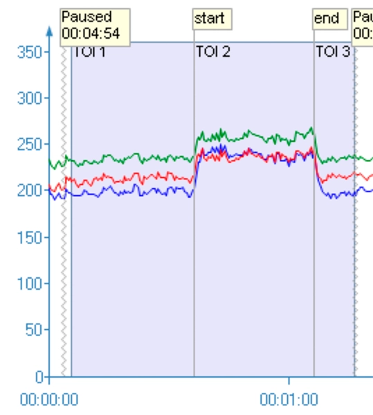
In addition to regions of interest you can create time periods of interest — TOIs. This feature is valuable for observing how blood flow dynamics evolve in response to stimuli, treatments, or other interventions. By setting TOIs in PIMSoft, you can capture and analyze perfusion data at key moments, such as baseline and stroke, enabling you to identify and quantify immediate and delayed physiological effects.
Perfusion overlay
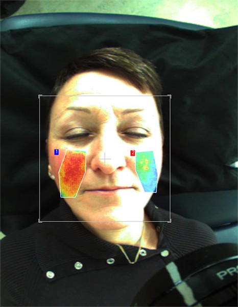
This feature enables perfusion images to be superimposed onto real images of the subject being studied, creating a precise visual alignment of physiological data with anatomical landmarks. Designed to help researchers see changes in localized blood flow relative to specific anatomical structures, pinpoint where perfusion changes occur, and analyze these in the context of targeted tissues or regions.
Reports
The robust reporting functionality streamlines data analysis and documentation. With PIMSoft, you can automatically generate detailed reports that include key perfusion metrics, graphical data, and images of selected regions and times of interest.
Reports can be customized to highlight specific data, such as perfusion changes over time, responses to interventions, or comparisons between baseline and post-stimulation measurements. Selected data can be exported as a PDF or spreadsheet, making it easy to share findings with colleagues, integrate data into broader studies, or include visual evidence in publications. This functionality is designed to save time while ensuring comprehensive, accurate records of research outcomes.
Application areas
PeriCam PSI HR is a versatile tool for preclinical research, providing high-resolution, real-time tissue perfusion that is critical to the investigation of physiological responses and disease mechanisms. Its noninvasive, laser-based imaging technology allows researchers to capture and quantify blood flow changes in small animals with precision, making it ideal for studies on vascular health, neurovascular function, wound healing, and drug efficacy.

Contact Sales
Get in touch
If you would like to know more about PeriCam PSI HR and how it can help you in your preclinical research, send us your contact details so that we can put a local representation in touch with you.
LASCA and LSCI
Laser speckle contrast imaging (LSCI) is the innovative technology behind PeriCam PSI, offering non-contact, high-resolution perfusion imaging. The technique was first described by J.D. Briers and S. Webster in 1996 and referred to as laser speckle contrast analysis (LASCA). In later years, the term LSCI gained in popularity and is nowadays the more common term. However, LSCI and LASCA both refer to the same technique, providing precise and reliable perfusion data without the need for contrast agents.